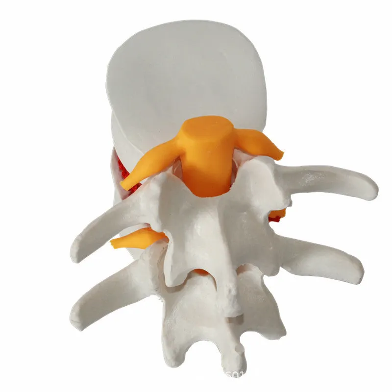
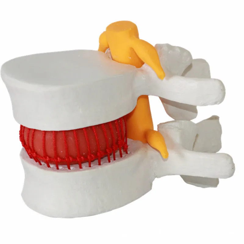
Medical educational model showing human lumbar intervertebral disc herniation anatomy for teaching and demonstration, made from PVC material measuring 11x10x7.6cm with removable disc components and white colour for clear visualisation.


Medical educational model showing human lumbar intervertebral disc herniation anatomy for teaching and demonstration, made from PVC material measuring 11x10x7.6cm with removable disc components and white colour for clear visualisation.
Medical anatomical model demonstrating lumbar intervertebral disc herniation for education

This PVC anatomical model provides healthcare educators, medical students, and clinicians with a tangible three-dimensional representation of lumbar intervertebral disc herniation. Measuring 11x10x7.6cm, the compact design makes it suitable for classroom demonstrations, patient consultations, or individual study. The white colour scheme enhances visual clarity, allowing detailed examination of spinal structures and pathological changes associated with disc herniation. Unlike flat diagrams or digital images, this physical model offers hands-on learning opportunities that improve anatomical understanding and clinical correlation.
Features and Construction
The model's construction focuses on accurate anatomical representation within practical dimensions for educational use. Each component is designed to demonstrate specific aspects of spinal pathology while maintaining durability for repeated handling.
Material and Build
Constructed from PVC plastic, the model balances detailed reproduction with structural stability. The material allows for precise moulding of anatomical features while withstanding regular handling in educational environments. The white colour provides optimal contrast for visualising spinal structures, making pathological changes more apparent during demonstrations. The removable disc components enable instructors to show both normal anatomy and herniated conditions.
Size and Practical Fit
With dimensions of 11x10x7.6cm, the model fits comfortably on desks, demonstration tables, or consultation room surfaces. The compact size facilitates easy transport between teaching locations while maintaining sufficient scale for clear visualisation. At 0.22kg, the lightweight design allows for one-handed demonstration during lectures or patient consultations. The packaging measures 11.5x11x8cm, providing protective storage without excessive bulk.
Uses and Placement

This anatomical model serves multiple educational functions across healthcare training and clinical environments. Its specific design addresses common learning challenges in understanding spinal pathology.
Event or Professional Use
In formal educational settings, the model supports anatomy and pathology lectures for medical, nursing, physiotherapy, and chiropractic students. Clinical educators use it to demonstrate disc herniation mechanics during patient education sessions, helping individuals visualise their condition. Professional development workshops benefit from the tangible representation when teaching spinal assessment techniques or discussing treatment options with colleagues.
Everyday Home Use
For self-directed learners, the model provides a study aid for anatomy revision outside formal classroom environments. Healthcare students can use it for personal examination of spinal structures, complementing textbook learning. Clinical practitioners may keep it in consultation rooms for occasional patient demonstrations when explaining diagnostic findings or treatment plans.
Benefits and Buying Value

The model addresses specific educational needs while offering practical advantages over alternative teaching tools. Its design prioritises clarity and durability within its intended applications.
Reuse and Low Maintenance
The PVC construction withstands repeated handling without significant wear, maintaining visual clarity across multiple teaching sessions. The material requires only occasional cleaning with a soft cloth to remove fingerprints or dust accumulation. The compact packaging protects components during storage or transport, extending the model's usable lifespan in educational settings where resources may be shared between departments or courses.
Why Choose This Product
This model provides a physical representation of disc herniation that enhances understanding beyond textbook diagrams or digital images. The specific focus on lumbar anatomy addresses a common area of spinal pathology relevant to multiple healthcare disciplines. The combination of removable components, clear visual design, and practical size makes it suitable for various teaching scenarios without requiring specialised display equipment or excessive storage space.